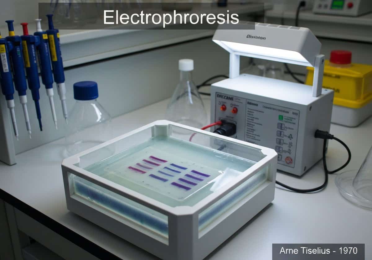
Gel electrophoresis is a cornerstone technique in molecular biology for the separation and analysis of macromolecules, primarily DNA, RNA, and proteins. The principle behind the technique is the migration of charged molecules through a porous gel matrix under the influence of an electric field. Since nucleic acids (DNA and RNA) have a consistently negative charge due to their phosphate backbone, they will naturally migrate towards the positive electrode (anode) when a current is applied. The gel itself, typically made of agarose for larger molecules like genomic DNA fragments or polyacrylamide for smaller fragments and proteins (in a technique called PAGE), acts as a molecular sieve. The matrix is composed of a network of pores through which the molecules must travel. Smaller molecules navigate this maze more easily and quickly than larger molecules, which are impeded more by the gel matrix. Consequently, the molecules are separated based on their size, with the smallest traveling the farthest from the starting point (the well) in a given amount of time. To perform DNA electrophoresis, a sample is first loaded into wells at one end of the gel. A ‘DNA ladder,’ a mixture of DNA fragments of known sizes, is run in a separate lane to serve as a reference for estimating the size of the unknown fragments. After the electric current is applied for a period, the separated fragments are visualized. Because DNA is invisible to the naked eye, a fluorescent dye that intercalates with the DNA, such as ethidium bromide or SYBR Green, is used. When placed under UV light, the dye fluoresces, revealing the DNA as distinct bands, each band representing a collection of fragments of the same size.